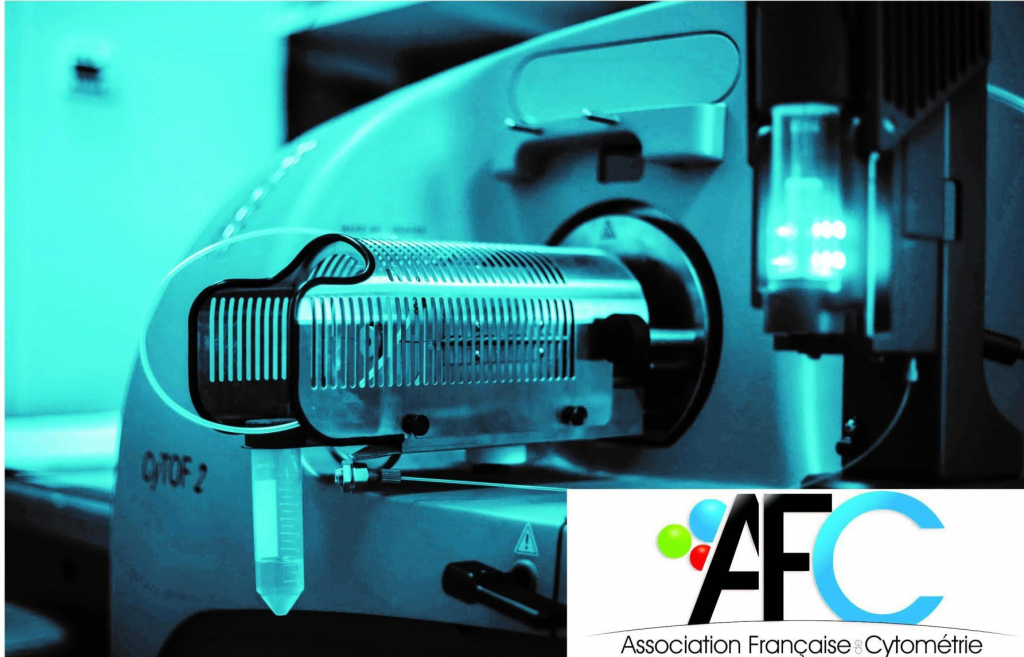
Thematic Group Webinars

Groupe Cytométrie de masse AFC : IL-12 and IL-23: paradoxical behavior in inflammation and cancer

Le groupe de travail “Cytométrie de masse” de l’Association Française de Cytométrie est heureux de vous inviter à son prochain webinaire :

Présenté par Pr. Burkhard Becher

University of Zurich, Institute of Experimental Immunology

Lien Zoom: https://univ-amu-fr.zoom.us/j/81537671135?pwd=VzJkTUtFdWVTMm0weUZvaVE1NTE3dz09

Code : 275002

IL-12 and IL-23 are closely related cytokines and represent the most proinflammatory mediators of the IL-12 superfamily. Both cytokines are structurally similar and share both producing and sensing cell types. While IL-12 is a canonical TH type I inducing mediator, IL-23 is firmly linked with type III immunity. Both cytokines are considered central mediators of TH cell polarization and are linked with inflammation and even immunopathology. However, whilst IL-12 stimulates type 1 lymphocytes and can eliminate established tumors, the loss of IL-12 leads to drastically enhanced inflammation across various models of chronic inflammatory disease and autoimmunity. Likewise, IL-23, which plays a non-redundant function in the development of autoimmunity and type 3 immune responses, it also increases tumor growth. These at first glance paradoxically appearing properties form the focus of this presentation. I will discuss the discoveries around IL-12 and IL-23 from a historical perspective, taking into account the most recent findings on their immunoregulatory properties in the context of inflammation and cancer in particular and immunity in general.

Mass Cytometry group AFC : Characterization of Regulatory T Lymphocytes in Breast Cancers

The Mass Cytometry Group of the French Cytometry Association Invites you to its Webinar

Tuesday November 7, 2023 at 12:30 p.m

Characterization of Regulatory T Lymphocytes in Breast Cancer

Presented by Stephane Fattori

Immunomonitoring department, Pauli Calmettes Institute, CRM, Marseille

Zoom link : https://univ amu fr.zoom.us/j/86931336119?pwd= TEgzb01ZM0FucFhKb3hEdmk5VEpKZz09
Login code: 725552

Abstract :
Triple negative breast cancer (TNBC) is the deadliest subtype of breast cancer Standard treatments for
patients with early stage, locally advanced or metastatic TNBC include the combination of neoadjuvant
chemotherapy and pembrolizumab, an immune checkpoint blockade targeting the programmed cell
death 1 (PD-1) receptor PD-1 blockade reinvigorate tumor antigen specific CD8+ T cells in primary TNBC.
However, approximately 40 and 80 of early stage and advanced or metastatic TNBC are resistant to
PD 1 blockade therapy, respectively.
Regulatory T cells (Tregs) are an immunosuppressive subset of CD4+ T cells essential for maintaining self
tolerance in physiological settings. Abundant tumor infiltration by Tregs harbouring hallmarks of highly
suppressive CD25high effector Tregs (eTregs) has been associated with resistance to PD 1 blockade in
cancer indications for which pembrolizumab was approved much earlier than for TNBC, such as non
small cell lung cancer. In contrast, in TNBC, the prognostic value of intratumoral Tregs remains
controversial and no association between baseline levels of eTregs and responses to PD 1 blockade
therapy has been reported, despite the clinical significance of this cancer indication. In the present
thesis, we used single cell technologies to dissect the nature and function of eTreg during TNBC
progression. We found that an immunosuppressive TNBC microenvironment resistant to PD-1 blockade
therapy is marked by an imbalance between effector CD8+ T cells and CD25 high eTregs. We further
identified CD25 as the most selective surface marker of eTregs in primary TNBC and associated
metastases for limited on target/off target effects. In a syngeneic TNBC model, selective Treg depletion
by a next generation anti-CD25 monoclonal antibody (mAb; ALD 2510; Alderaan Biotechnology)
synergized with PD-1 blockade to safely mediate durable tumor growth control by increasing effector
αβCD8+ T cell/Treg ratios in tumors and in the peripheral blood Overall, we propose that Treg depletion
by next generation anti-CD25 mAb could improve clinical responses to PD-1 blockade therapy in patients
with TNBC at different stage of the disease

Data Analysis : Build an informative abundance heatmap.

The Data Analysis Group of the French Cytometry Association

Invites you to its 3nd Webinar 2023

Decryption

Build an informative abundance heatmap.

Presented by :

S. Granjeaud

Plateforme Bioinformatique

Centre de Recherche en Cancérologie de Marseille, Marseille, France

Thursday June 29, 2023 / 13h00 – 14h00

Ask your questions on the forum of AFC before the webinar
Login link ZOOM at the webinar

Please spread this announcement around you.


See you soon 

Aïda MEGHRAOUI & Samuel GRANJEAUD 

Find us on the group page

Groupe Cytométrie de masse AFC : Live-Cell Barcoding with Anti-CD45 Antibodies for Mass Cytometry

Le groupe de travail “Cytométrie de masse” de l’Association Française de Cytométrie est heureux de vous inviter à son prochain webinar :

Mercredi 7 juin à 13h

Live-Cell Barcoding with Anti-CD45 Antibodies for Mass Cytometry

Présenté par Pauline Maby

Immunomonitoring department, Pauli Calmettes Institute, Marseille

Lien zoom : https://univ-amu-fr.zoom.us/j/87588252730?pwd=ek5ZQytnSEd6Q21ZM3FvZ3Bka3lQZz09

Résumé : Le codage à barres CD45 des cellules vivantes implique la coloration d’échantillons de cellules immunitaires avec des combinaisons uniques d’anticorps anti-CD45 comme identificateurs d’échantillons. Cela permet le regroupement d’échantillons afin de les traiter comme un échantillon de cellule unique pour la coloration et l’acquisition des cellules. Il améliore le flux de travail, améliore la cohérence des données et réduit l’utilisation de réactifs.

Au cours de ce webinaire, nous discuterons de toutes les étapes d’une expérience de codage à barres CD45 sur cellules vivantes, de la conception du panel à l’analyse des données. Tout d’abord, lors du panel design, il est important de s’assurer que la coloration CD45 n’interfère pas avec la détection de marqueurs d’intérêt dans d’autres canaux. Ensuite, plusieurs ajustements doivent être apportés aux procédures de coloration et d’acquisition. Enfin, les fichiers FCS obtenus à partir d’échantillons de cellules à code-barres sont décodés pour attribuer chaque événement cellulaire à son échantillon d’origine.

En espérant vous y retrouver nombreux !

A très bientôt ! 

Olivier MOLENDI-COSTE & Anne-Sophie CHRETIEN

Nous retrouver sur la page du groupe


GT Microbiologie-Cytométrie : Vitalité cellulaire

Le Groupe Microbiologie-Cytométrie de l’Association Française de Cytométrie

Vous invite à son Webinar 2023

Vitalité Cellulaire

La vitalité cellulaire dépend de la charge énergétique des cellules. Elle se traduit par la cinétique des réactions métaboliques ou activité métabolique. Dans les industries de fermentation, les ferments doivent être viables mais aussi actifs pour coloniser rapidement le milieu en début de fermentation. On peut aussi évaluer l’effet de stress sur les cellules par la mesure de vitalité. La cytométrie en flux permet de mesurer en 30min la vitalité cellulaire, ce qui est plus simple et plus rapide que les mesures cinétiques d’activités métaboliques. Ce webinaire présentera la méthode avec quelques exemples de corrélation avec des mesures d’activités sur des levures et des bactéries.

Présenté par :

Marielle BOUIX

Responsable du groupe thématique Microbiologie-Cytométrie SFM

Mardi 30 Mai 2023 / 13h30 – 14h15


Lien de connexion au webinar : Cliquez

Merci de diffuser cette annonce autour de vous.


A très bientôt ! 

Société Française de Microbiologie

36 avenue Jean Moulin 75014 Paris

Tel.+339-63-04-70-73

secretariat@sfm-microbiologie.org / comptabilite@sfm-microbiologie.org

www.sfm-microbiologie.org

Nous retrouver sur la page du groupe

Data Analysis: Decryption, to read a heatmap, you have to know how to build it.

The Data Analysis Group of the French Cytometry Association

Invites you to its 2nd Webinar 2023

Decryption

To read a heatmap you have to know how to build it

Presented by :

S. Granjeaud

Plateforme Bioinformatique

Centre de Recherche en Cancérologie de Marseille, Marseille, France

wednesday may 10, 2023 / 13h00 – 14h00

Ask your questions on the forum of AFC before the webinar
Login link ZOOM at the webinar

Please spread this announcement around you.


See you soon 

Aïda MEGHRAOUI & Samuel GRANJEAUD 

Find us on the group page

Mass Cytometry : Enhanced single-cell segmentation and whole slide imaging analysis through dual-modality immunofluorescen

The Mass Cytometry Group of the French Cytometry Association Invites you to its Webinar

Enhanced single-cell segmentation and whole slide imaging analysis through dual-modality immunofluorescence and imagingmass cytometry  (IMC)

Presented by : John Lizhe Zhuang

Department of Oncology, University of Cambridge

Tuesday may 2, 2023 at 16h00

login link ZOOM

Please spread this announcement around you.


See you soon 

Olivier MOLENDI-COSTE & Anne-Sophie CHRETIEN

Find us on the group page

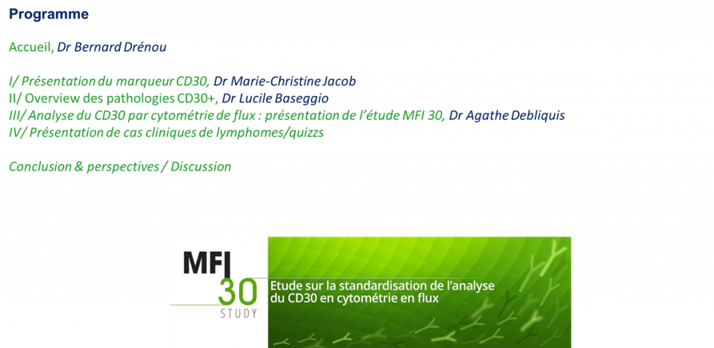
CYTHEM : all about CD30

Dear colleagues,

CytHem is pleased to inform you that, following the MFI30 cytometry work carried out under the aegis of the AFC, a webinar entitled “All about CD30” will take place on Friday October 21 from 1 p.m. to 2:30 p.m. (program below ).

Here is the Teams link to open with Chrome :

Click here to take part to a meeting

Hope to see many of you there !

The team ‘Cythem’

Image Cytometry : Cell Cycle in Image Cytometry

The Image Cytometry Working Group of the French Association of Cytometry offers a Webinar

Monday, October 3, 2022 from 1:30 p.m. to 2:15 p.m.

Interest of image cytometry in the evaluation of the expression and localization of the TRIM37 protein during the cell cycle in a chondrocyte line

Benjamin Brigant

Centre of Molecular Inflammation Research (CEMIR),  Department of Clinical and Molecular Medicine (IKOM),NTNU, Trondheim, Norvège

Registration is mandatory via the following form:  link

Registration deadline September 28, 2022. The meeting link will be sent to you once registration has been completed.

Hope to see many of you there!

The Image Cytometry group ’

V. Duplan-Eche, S. Dussurgey, Y. Demont, M. Dussiot, C. Guérin, J. Cosette, M. Boyer-Clavel, F. Hédin, ML.  Renoud, AL. Raveu, A. Malfait-Jobart  

contact : valerie.duplan@inserm.fr

Image Cytometry: Colocalization in Image Cytometry

The Image Cytometry Working Group of the French Association of Cytometry offers a Webinar

Monday March 21, 2022 from 1:30 p.m. to 2:45 p.m

  • 1:30-2:40 p.m. : Molecular and morphological polarizations in the T cells Jérôme Delon, Institut Cochin, Paris
  • 2:00-2:45 p.m. : Round table : Analysis data on collocation (IDEAS Software) Michaël Dussiot, Institut Imagine, paris

Registration is mandatory via the following form :  link

Registration deadline March 09, 2022. The meeting link will be sent to you nce you have registered.

Hope to see many of you there!

‘The Image Cytometry group’

V. Duplan-Eche, S. Dussurgey, Y. Demont, C. Guérin, M. Dussiot, J. Cosette, F. Hédin, T. Andrieu, M. Boyer-Clavel

contact : valerie.duplan@inserm.fr